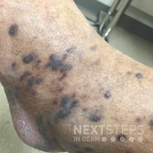
The correct answer is B. HHV-8.
The figure illustrates lesions of Kaposi sarcoma (KS), which typically manifest as blue-black to violaceous macules, papules, or nodules. KS lesions stain positively for HHV-8 which is thought to be involved in the pathogenesis.
HPV may be used to identify viral warts.
PAS (periodic acid Schiff) stain is used to identify fungal organisms.
S100 is a melanocytic stain that can be used to identify melanoma and neuroendocrine tumors. CD207, or Langerin, stains Langerhans cells.
References:
Reference: Buonaguro FM et al. Kaposi’s sarcoma: Aetiopathogenesis, histology and clinical features. J Eur Acad Dermatol Venereol. 2003; 17: 138-54.
