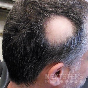
The correct answer is D. Peribulbar lymphocytic inflammation, dilated infundibula, and increased telogen count.
The image, along with the vignette, describes Alopecia Areata (AA) (see image Figure 2.5.1 in DIR). AA can present as a localized area (in this case), the entire scalp (totalis), or the whole body (universalis). On physical exam, the scalp skin looks normal with “exclamation point” hair (tapering at proximal end) and nail pits can also be seen. Some autoimmune diseases can often be associated with AA including thyroid disease, vitiligo, diabetes, systemic lupus erythematosus, and myasthenia gravis. Biopsy shows peribulbar lymphocytic inflammation (“swarm of bees”), dilated infundibula, and increased telogen count (see Table 5.10.1). Biopsy technique is important when evaluating alopecia. For nonscarring conditions, obtain two 4mm punch biopsies (one from unaffected area and one from affected region). An interesting fact about AA is that it targets mostly pigmented hair, giving the illusion of an increased number of white hairs with diffuse AA.
1. Distorted follicular anatomy, perifollicular hemorrhage, and pigment casts are histopathologic findings seen in trichotillomania (see Table 5.10.1) 2. Spongiotic or psoriasiform epidermis, atrophic sebaceous glands, markedly elevated telogen count, and increased miniaturization are histopathologic findings seen in psoriatic alopecia (see Table 5.10.1) 3. Peribulbar inflammation with plasma cells, increased miniaturization, and increased telogen count are histopathologic findings seen in syphilitic alopecia (see Table 5.10.1) 4. Peribulbar lymphocytic inflammation, dilated infundibula, and increased telogen count is the correct answer 5. Periadnexal lymphocytes, vacuolar interface, and increased mucin are histopathologic findings seen in non-scarring alopecia of systemic lupus (see Table 5.10.1)
References: Medical Dermatology chapter 2.5 Disorders of Hair and Nails, DIR Review book page 149; Dermatopathology chapter, 5.10 Alopecia, DIR Review book page 353 2021
