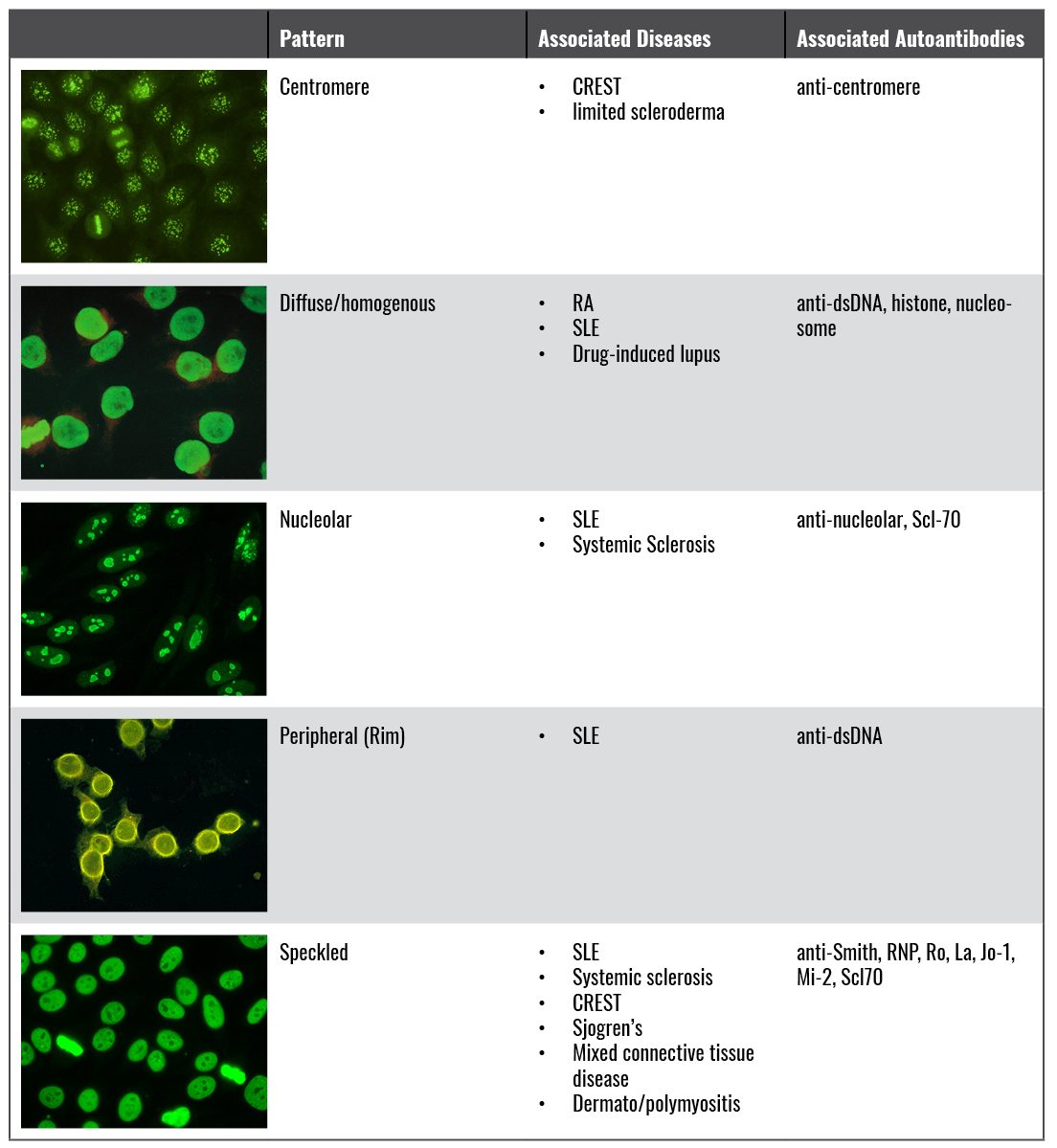
Connective tissue diseases are a boards favorite, and lab interpretation, including identifying ANA patterns from photos, are fair game. I’ve put together the following table of ANA patterns as well as their associated disease states and autoantibodies for easy memorization (and easy points!).
Image sources:
Centromere: https://www.researchgate.net/figure/Centromere-staining_fig9_45281682
Diffuse: http://www.mbl.co.jp/e/ivd/atlas_pattern.html
Nucleolar: https://commons.wikimedia.org/wiki/File:ANA_NUCLEOLAR_3.jpg
Peripheral: https://library.med.utah.edu/WebPath/IMMHTML/IMM003.html


