
Using Dermoscopy to Diagnose Difficult Clinical Conditions
940594059405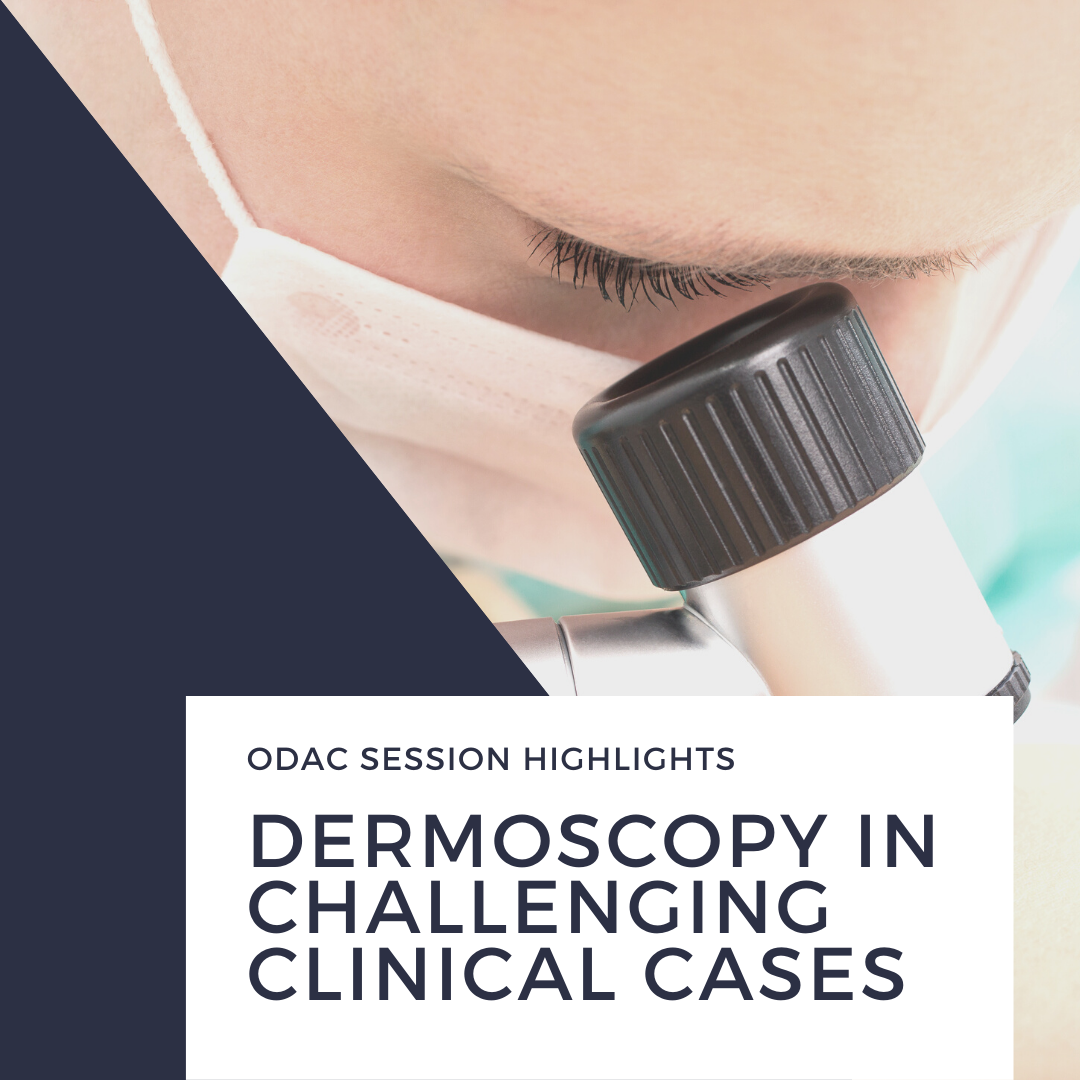 Poised at the microphone, Dr. Sima Jain taught us how to use dermoscopy to diagnose difficult clinical cases during the 17th Annual ODAC Dermatology, Aesthetic and Surgical Conference in Orlando, Florida. Dermoscopy uses epiluminescent microscopy to help the provider get a more in-depth examination of the skin. It is extremely helpful for aiding the diagnosis of pigmented skin lesion …
Poised at the microphone, Dr. Sima Jain taught us how to use dermoscopy to diagnose difficult clinical cases during the 17th Annual ODAC Dermatology, Aesthetic and Surgical Conference in Orlando, Florida. Dermoscopy uses epiluminescent microscopy to help the provider get a more in-depth examination of the skin. It is extremely helpful for aiding the diagnosis of pigmented skin lesion … Continue reading "Using Dermoscopy to Diagnose Difficult Clinical Conditions"
 Next Steps in Derm author, Dr. Anna Chacon, searched the journals so that you don’t have to! She reports on important take-aways from different dermatology journals for the months of January, February, and March of 2019.
It is key to keep in mind that “important” is subjective and what is contained in this review is one person’s view of what should be remembered from these months of the …
Next Steps in Derm author, Dr. Anna Chacon, searched the journals so that you don’t have to! She reports on important take-aways from different dermatology journals for the months of January, February, and March of 2019.
It is key to keep in mind that “important” is subjective and what is contained in this review is one person’s view of what should be remembered from these months of the … 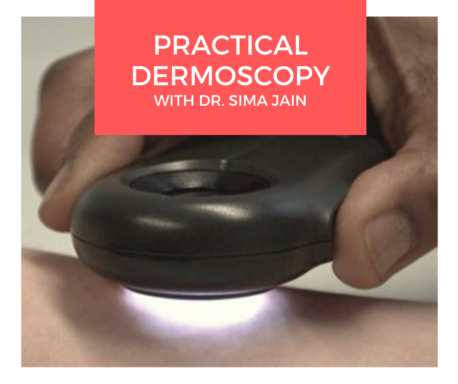 Dermoscopy, also known as epiluminescence microscopy, epiluminoscopy or skin surface microscopy, is an important way to visualize subsurface structures in the epidermis and dermis. In a 2-part series, Dr. Sima Jain reviews the evaluation of pigmented lesions, and the different vessel morphologies and patterns along with a discussion of specific findings in select cutaneous infections.
Read part …
Dermoscopy, also known as epiluminescence microscopy, epiluminoscopy or skin surface microscopy, is an important way to visualize subsurface structures in the epidermis and dermis. In a 2-part series, Dr. Sima Jain reviews the evaluation of pigmented lesions, and the different vessel morphologies and patterns along with a discussion of specific findings in select cutaneous infections.
Read part … 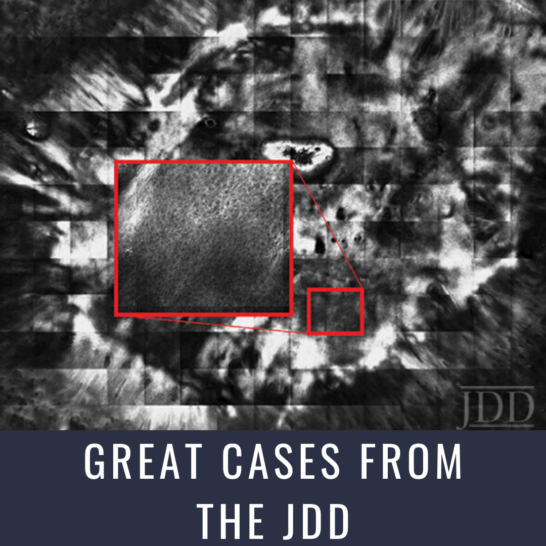 Reflectance Confocal Microscopy (RCM) is a new noninvasive skin imaging modality that is comparable to traditional histopathology. Authors Radhika Srivastava BA, Catherine Reilly BS, Gina Francisco MBS, Hamza Bhatti DO, and Babar K. Rao MD present serial in vivo RCM imaging of an atypical nevus after shave excision over a 1-month period. Findings on RCM images are consistent with the inflammatory, …
 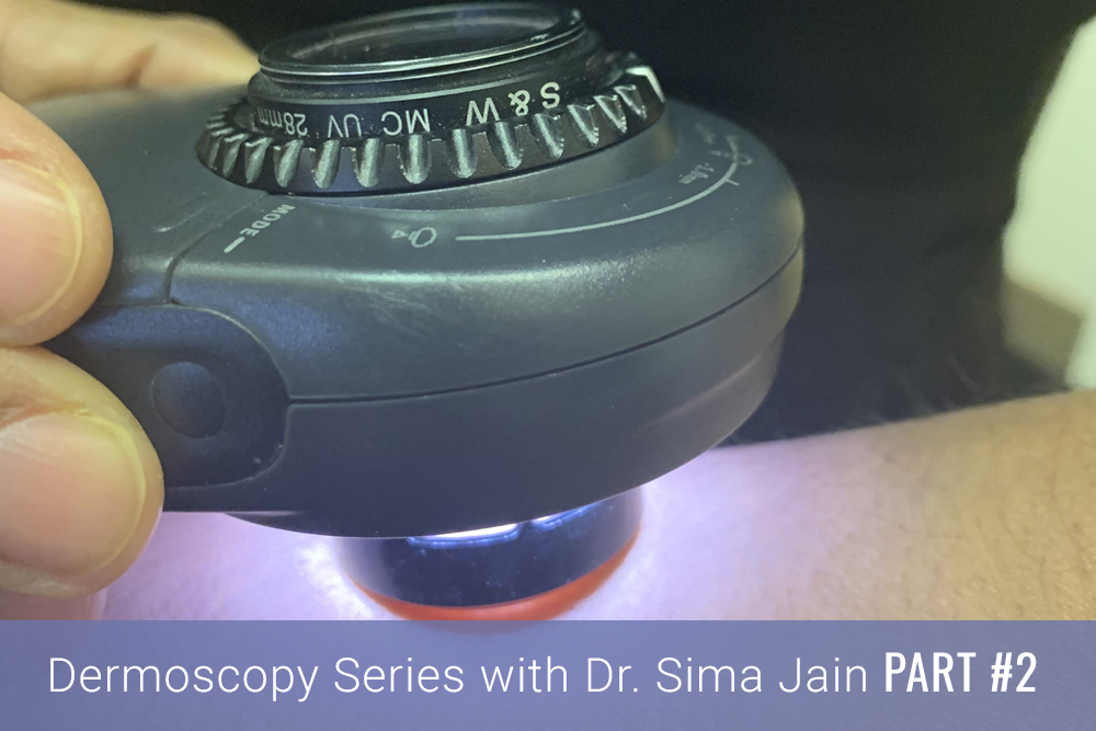 Introduction
Dermoscopy, also known as epiluminescence microscopy, epiluminoscopy or skin surface microscopy, is an important way to visualize subsurface structures in the epidermis and dermis. Part one of this article focused on the evaluation of pigmented lesions, and the second installment below will review the different vessel morphologies and patterns along with discussing specific findings …
Introduction
Dermoscopy, also known as epiluminescence microscopy, epiluminoscopy or skin surface microscopy, is an important way to visualize subsurface structures in the epidermis and dermis. Part one of this article focused on the evaluation of pigmented lesions, and the second installment below will review the different vessel morphologies and patterns along with discussing specific findings …