
Benefits & Pitfalls of Gene Expression Profiling in Treating Cutaneous Malignancies
152801528015280 Next Steps in Derm, in partnership with ODAC Dermatology, Aesthetic and Surgical Conference, interviewed Dr. Vishal A. Patel (fellowship trained Mohs micrographic surgeon who serves as Director of Cutaneous Oncology at the GW Cancer Center and Director of Dermatologic Surgery at the GW Department of Dermatology) about the pros and cons of gene expression profiling. Watch him describe how this tech …
Next Steps in Derm, in partnership with ODAC Dermatology, Aesthetic and Surgical Conference, interviewed Dr. Vishal A. Patel (fellowship trained Mohs micrographic surgeon who serves as Director of Cutaneous Oncology at the GW Cancer Center and Director of Dermatologic Surgery at the GW Department of Dermatology) about the pros and cons of gene expression profiling. Watch him describe how this tech … 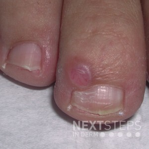 The mucin found in this lesion is produced by:
A. Fibroblasts
B. Keratinocyte
C. Merkel cells
D. Nail matrix
E. Osteoblast
To find out the correct answer and read the explanation, click here.
Brought to you by our brand partner Derm In-Review. A product of SanovaWorks.
…
The mucin found in this lesion is produced by:
A. Fibroblasts
B. Keratinocyte
C. Merkel cells
D. Nail matrix
E. Osteoblast
To find out the correct answer and read the explanation, click here.
Brought to you by our brand partner Derm In-Review. A product of SanovaWorks.
…  Histologically, this lesion shows plump, polygonal cells arranged in nests and fascicles with granular cytoplasm. Which immunohistochemical stain would be positive?
A. CD31
B. Synaptophysin
C. Factor XIIIa
D. S-100
E. CD34
To find out the correct answer and read the explanation, click here.
Brought to you by our brand partner Derm In-Review. A product of SanovaWorks.
…
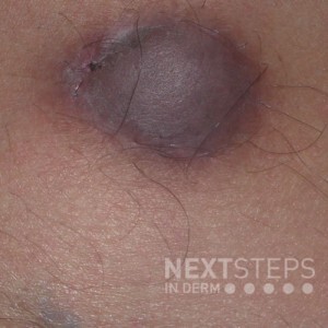
Histologically, this lesion shows plump, polygonal cells arranged in nests and fascicles with granular cytoplasm. Which immunohistochemical stain would be positive?
A. CD31
B. Synaptophysin
C. Factor XIIIa
D. S-100
E. CD34
To find out the correct answer and read the explanation, click here.
Brought to you by our brand partner Derm In-Review. A product of SanovaWorks.
…  Which of the following is the least likely cause of the below lesion?
A. Cryptococcosis
B. Histoplasmosis
C. Coccidiodomycosis
D. Penicilliosis
E. Lobomycosis
To find out the correct answer and read the explanation, click here.
Brought to you by our brand partner Derm In-Review. A product of SanovaWorks.
…
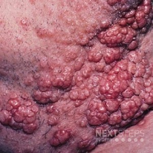
Which of the following is the least likely cause of the below lesion?
A. Cryptococcosis
B. Histoplasmosis
C. Coccidiodomycosis
D. Penicilliosis
E. Lobomycosis
To find out the correct answer and read the explanation, click here.
Brought to you by our brand partner Derm In-Review. A product of SanovaWorks.
…  What would this lesion show histologically?
A. A proliferation of basaloid cells with small ducts
B. A grenz zone with a lot of inflammatory cells and globi
C. Well formed tuberculoid granulomas in a linear pattern
D. Scattered comedo-like cysts
E. Acanthosis and neutrophilic infiltrate
To find out the correct answer and read the explanation, click here.
Brought to you by our …
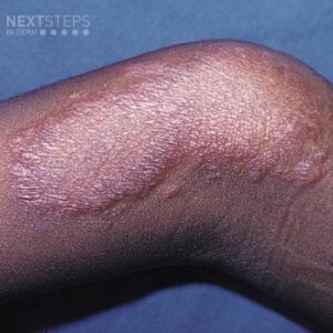
What would this lesion show histologically?
A. A proliferation of basaloid cells with small ducts
B. A grenz zone with a lot of inflammatory cells and globi
C. Well formed tuberculoid granulomas in a linear pattern
D. Scattered comedo-like cysts
E. Acanthosis and neutrophilic infiltrate
To find out the correct answer and read the explanation, click here.
Brought to you by our …