Using Lasers & EBDs for Pigmented Lesions & Tattoos
213902139021390 During the 2023 Skin of Color Update in New York City, Dr. Omar Ibrahimi, a renowned laser and cosmetic dermatologist, as well as a Mohs surgeon in private practice in Stamford, Connecticut, imparted valuable insights into the use of lasers for pigmented lesions and tattoos. Dr. Ibrahimi placed significant emphasis on ensuring both safety and efficacy, particularly in individuals with diverse skin …
 During the 2023 Skin of Color Update in New York City, Dr. Omar Ibrahimi, a renowned laser and cosmetic dermatologist, as well as a Mohs surgeon in private practice in Stamford, Connecticut, imparted valuable insights into the use of lasers for pigmented lesions and tattoos. Dr. Ibrahimi placed significant emphasis on ensuring both safety and efficacy, particularly in individuals with diverse skin …
 Continue reading "Using Lasers & EBDs for Pigmented Lesions & Tattoos"


 Admittedly, it took me a while to get over the fear of an artificial intelligence (AI) “apocalypse”, which likely developed after my older brother forced me to repeatedly watch “The Terminator” at the tender age of seven. Through an extensive dive into the literature and numerous lectures by Dr. Vishal A. Patel, I’ve since realized the applicability and patient benefit of AI in dermatolo …
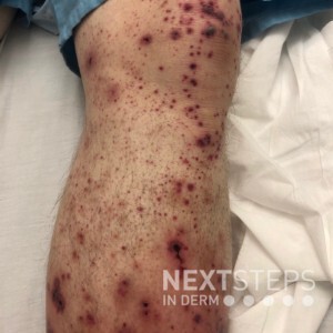
  Similar lesions are present on the contralateral leg, and this patient also reports joint pain and tarry stools. A biopsy for direct immunofluorescence is most likely to show which of the following?
A. IgA deposition in the dermal papillae
B. Perivascular IgA deposition
C. Perivascular IgG deposition
D. Linear IgA deposition along the basement membrane zone
E. Linear IgG deposit …
  Dermoscopy, also known as epiluminescence microscopy, epiluminoscopy or skin surface microscopy, is an important way to visualize subsurface structures in the epidermis and dermis. In a 2-part series, Dr. Sima Jain reviews the evaluation of pigmented lesions, and the different vessel morphologies and patterns along with a discussion of specific findings in select cutaneous infections.
Read part …
  Introduction
Dermoscopy, also known as epiluminescence microscopy, epiluminoscopy or skin surface microscopy, is an important way to visualize subsurface structures in the epidermis and dermis. Part one of this article focused on the evaluation of pigmented lesions, and the second installment below will review the different vessel morphologies and patterns along with discussing specific findings …